
Athletes and Right Ventricular Dysfunction
Historically, the athlete's heart was first described in 1899 by Swedish physician S. Henschen. Using percussion and auscultation, he observed that Northern European skiers had larger hearts than individuals leading sedentary lifestyles. That same year, E. Darling conducted similar studies on rowers at Harvard University. In 1900, P. D. White examined long-distance runners participating in the Boston Marathon and identified sinus bradycardia (1).
Interest in athletes and their cardiovascular adaptations remains considerable today. Consequently, researchers continue to seek the latest and most advanced diagnostic methods to investigate changes in the myocardium, the muscular tissue of the heart, resulting from intense physical exercise.
Metabolic Equivalents of Task
Metabolic equivalents of task (METs) are used to objectively assess physical exertion. Table 1 illustrates the intensity of various physical activities and their corresponding MET values.
Table 1. Physical Activity and Corresponding MET Values
| Physical Activity Intensity | Activities and MET Values |
|---|---|
| Light physical activity (MET < 3) | • Sleeping (0.9) |
| • Watching television (1.0), writing (1.8) | |
| • Slow walking at 2.7 km/h (2.3) | |
| • Walking at 4 km/h (2.9) | |
| Moderate physical activity (MET 3–6) | • Cycling (3.0–5.5) |
| • Brisk walking (3.3–3.6) | |
| • Jumping (4.8) | |
| Vigorous physical activity (MET > 6) | • Running (11.0) |
| • Gymnastics (8.0) | |
| • Basketball (8.0) | |
| • Swimming (8.0) |
One MET is equivalent to oxygen consumption at rest: 1 MET = 3.5 mL O₂/kg/min. According to the World Health Organization (WHO), 42–70 METs per week are recommended for all healthy individuals to reduce the risk of cardiovascular disease (CVD). In contrast, athletes' weekly physical activity levels are 5–6 times higher, reaching up to 200–300 METs per week.
Study Results
When discussing the athlete's heart, attention is most often focused on physiological and structural changes in the left ventricle (LV). However, various studies have demonstrated that changes in the right ventricle (RV) are also significant. Research indicates that high-endurance exercise induces RV remodeling, and these changes are not always reversible (2, 3).
Over time, myocardial inflammation and fibrosis may lead to the formation of a proarrhythmic substrate and myocardial dysfunction, particularly in athletes with a genetic predisposition (4). RV arrhythmias and chronic RV remodeling have most frequently been observed in highly trained athletes competing in long-distance events (5). Clinical studies have shown that more than 50% of marathon runners have elevated blood troponin I (TnI) concentrations (6).
One clinical study included 40 athletes. The main inclusion criteria required participants to be highly trained, defined as engaging in more than 10 hours of physical activity per week or having completed at least one-quarter of the course distance of their respective competitions. Participants also had to be asymptomatic, have no cardiovascular risk factors, and show no structural or electrophysiological abnormalities.
Of the participants, 7 were preparing to compete in a marathon (42.2 km), 11 in a triathlon (1.9/90/21.1 km), 9 in a mountain biking event (207 km), and 13 in an ultratriathlon (3.8/180/42.2 km).
Echocardiographic assessment of RV and LV parameters before and after competition (Tables 2 and 3) revealed significant RV changes following intense physical exertion.
Table 2. Baseline Echocardiographic Parameters
| Echocardiographic Measurements | All Participants | Marathon Runners | Triathletes | Mountain Bikers | Ultratriathletes | P-value |
|---|---|---|---|---|---|---|
| RV end-diastolic volume (mL) | 170±30 | 177±31 | 161±38 | 161±24 | 181±20 | 0.258 |
| LV end-diastolic volume (mL) | 150±23 | 159±11 | 143±26 | 142±21 | 157±26 | 0.237 |
| RV end-systolic volume (mL) | 83±17 | 85±17 | 80±21 | 80±15 | 90±12 | 0.410 |
| LV end-systolic volume (mL) | 66±14 | 66±5 | 65±16 | 60±13 | 71±15 | 0.241 |
| RVEF (%) | 51.0±3.6 | 51.9±3.1 | 50.75±5.0 | 50.5±4.0 | 50.3±3.2 | 0.849 |
| LVEF (%) | 56.4±5.2 | 58.2±2.6 | 55.0±5.6 | 58.4±5.8 | 54.8±5.1 | 0.239 |
| TAPSE (mm) | 11.0±1.5 | 11.1±1.1 | 10.5±0.9 | 10.9±1.8 | 11.8±1.5 | 0.225 |
| LV diastolic diameter (mm) | 56.0±4.9 | 54.5±4.8 | 54.1±3.8 | 57.9±4.5 | 57.7±5.4 | 0.162 |
| RV fractional shortening (%) | 51.5±6.0 | 50.7±5.2 | 51.3±6.0 | 52.6±5.3 | 51.5±7.3 | 0.942 |
| RV systolic strain (%) | −27.2±3.4 | −27.2±1.2 | −26.5±3.8 | −28.8±3.3 | −26.6±3.9 | 0.292 |
| LV systolic strain (%) | −18.4±3.7 | −18.0±3.6 | −17.5±3.8 | −19.8±3.8 | −17.8±2.7 | 0.469 |
Table 3. Changes in Parameters After Competition
| Echocardiographic Parameters | Baseline | Immediately After Competition | After Competition | P-value |
|---|---|---|---|---|
| Systolic BP (mmHg) | 147±14 | 117±13 | 134±20 | <0.0001 |
| Diastolic BP (mmHg) | 77±7 | 70±11 | 74±10 | 0.001 |
| Pulse pressure (mmHg) | 21.5±3.8 | 18.0±3.3 | 20.0±3.3 | <0.0001 |
| RVEF (%) | 51.0±3.6 | 46.4±6.5 | 50.0±3.8 | <0.0001 |
| RV fractional shortening (%) | 51.5±6.0 | 44.3±11.2 | 49.8±6.6 | <0.0001 |
| RV systolic strain (%) | −27.2±3.4 | −23.7±3.7 | −25.6±3.0 | 0.001 |
| RV systolic strain rate (s⁻¹) | −1.42±0.24 | −1.26±0.23 | −1.29±0.19 | 0.008 |
| LVEF (%) | 56.4±5.2 | 57.5±5.6 | 58.8±5.1 | 0.147 |
| LV systolic strain (%) | −18.4±3.7 | −16.9±2.8 | −17.7±2.3 | 0.071 |
| LV systolic strain rate (s⁻¹) | −0.98±0.26 | −0.95±0.15 | −0.89±0.13 | 0.13 |
Abbreviations: RV – right ventricle; LV – left ventricle; RVEF – right ventricular ejection fraction; LVEF – left ventricular ejection fraction; TAPSE – tricuspid annular plane systolic excursion; BP – blood pressure.
The mean age of the athletes was 37 ± 8 years, and 90% were male. Their average weekly training duration was 16.3 ± 5.1 hours. The athletes underwent three assessments: 2–3 weeks before competition, immediately after competition (approximately 20–40 minutes afterward), and 6–11 days after competition.
The results (Table 3) showed that all echocardiographic parameters of the right ventricle (RV), including right ventricular ejection fraction (RVEF), RV fractional shortening, systolic strain, and systolic strain rate, decreased significantly after competition. In contrast, left ventricular (LV) parameters remained unchanged.
The reduction in RV function was also directly associated with the duration of the event. The greatest decrease in RVEF was observed among ultratriathletes, whose events lasted approximately 11 hours, while the smallest reductions occurred among marathon runners (3 hours) and triathletes (5.5 hours) (Figure 1).
The athletes' biochemical markers were also evaluated. Elevated troponin I (TnI) levels were detected in 9 athletes (23%) before competition (mean 0.015 mg/L, range 0.015–0.21 mg/L) and in all athletes after competition (mean 0.018 mg/L, range 0.020–0.99 mg/L).
B-type natriuretic peptide (BNP) levels were also elevated after competition compared with baseline (13.1 ± 14.0 vs. 25.4 ± 21.4 ng/L, p = 0.003).
A stronger association between reduced RVEF and increased biomarker levels was observed among ultratriathletes (r = 0.746, p = 0.003 for TnI; r = 0.625, p = 0.022 for BNP). In contrast, no association was found between left ventricular ejection fraction (LVEF) and TnI (r = −0.065, p = 0.702) or BNP (r = 0.250, p = 0.125).
Late gadolinium enhancement (LGE) was detected in five athletes (Figure 2). These athletes underwent further evaluation, which revealed several distinguishing characteristics compared with athletes without LGE:
- They had participated in competitive sports for a longer period (20 ± 16 vs. 8 ± 6 years, p = 0.043).
- They had a higher age-adjusted predicted maximal oxygen uptake (VO₂max) (162 ± 26% vs. 144 ± 16%, p = 0.036).
- Their RV end-diastolic volume was 16% greater, and their RV end-systolic volume was 25% greater (p = 0.029 and p = 0.003, respectively).
- Their RVEF was lower (47.1 ± 5.9% vs. 51.1 ± 3.7%, p = 0.042).
- Their LVEF was similar to that of athletes without LGE (56.5 ± 6.8% vs. 59.8 ± 5.6%, p = 0.242).
Figure 2. Late gadolinium enhancement (LGE) was detected in five athletes. The arrows indicate focal areas of LGE, most prominent in the interventricular septum.
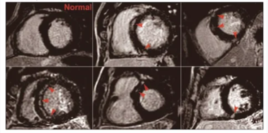
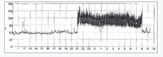
Figure 3. A 24-hour ECG recording showing an episode of paroxysmal ventricular tachycardia (VT) during the night.
Lone Atrial Fibrillation
Numerous clinical studies have investigated the association between prolonged, high-intensity endurance exercise and the development of atrial fibrillation or atrial flutter (AF/AFL). The first clinical studies were conducted in 1998 by Karjalainen and colleagues. A 10-year Scandinavian study involving orienteering athletes found that AF developed in 5.3% of athletes, compared with 0.9% in the control group. AF was more frequently diagnosed in middle-aged athletes without identifiable etiological factors.
In another study, Baldesberger and colleagues compared cyclists participating in the Tour de Suisse with golfers and found that cyclists had a 10% higher risk of developing AF/AFL (p = 0.028).
Frustaci and colleagues conducted a study involving 12 athletes with lone AF, a condition accounting for 2–10% of cases in the general population and diagnosed in individuals aged 60 years or younger without identifiable etiological risk factors for AF/AFL.
Histological examination revealed myocardial abnormalities, including inflammatory infiltration (66%), noninflammatory cardiomyopathy (17%), and fibrotic changes (17%) (7). These abnormalities may have contributed to the formation of a proarrhythmic substrate.
A typical clinical presentation might involve a middle-aged man (approximately 40–50 years old) who has participated in competitive sports, such as swimming, running, or cycling, since adolescence. Episodes of paroxysmal AF usually occur at night, resolve spontaneously, and almost never develop during physical activity (Figure 3).
Athletes generally continue their usual activities until the episodes become more frequent and AF progresses to a chronic form. In a study by Hoogsteen and colleagues, the incidence of such cases was reported to be as high as 17%.
Summary
In conclusion, endurance exercise causes acute right ventricular (RV) dysfunction in athletes, with the severity of dysfunction increasing according to the duration of physical exertion.
Repeated exposure to intense exercise may eventually lead not only to myocardial inflammation and fibrosis but also to an increased risk of cardiac rhythm disturbances. Accordingly, RV remodeling and late gadolinium enhancement (LGE) were more common among athletes with a longer history of competitive sports participation.
Cardiologist Viktorija Šiurkaitė
Lithuanian University of Health Sciences
