
How to Protect Your Brain From the Harmful Effects of High Blood Pressure
Introduction
Hypertension is one of the main health problems in developed countries. It is diagnosed when systolic blood pressure is higher than 140 mm Hg and diastolic blood pressure is higher than 90 mm Hg. According to statistical data, increased blood pressure affects more than 25% of the general population. It has been established that hypertension is one of the main risk factors for brain, heart, blood vessel, and kidney pathologies (1). Although there is a considerable amount of knowledge about the mechanisms of blood pressure control, the exact cause of hypertension is determined only for a small percentage of patients. When the causes of high blood pressure cannot be identified, primary or essential hypertension is diagnosed.
Currently, there are many effective medications on the market that help correct high blood pressure and reduce its negative impact on various organs and tissues. However, most of these drugs act systemically and are not organ-specific. In recent years, more attention has been paid to analyzing the impact of hypertension on specific organs. This helps to better understand how and what kind of damage can develop in the body due to high blood pressure and to select the most effective organ-targeted protective measures (1).
One of the main organs negatively affected by high blood pressure is the brain. Hypertension significantly increases the risk of stroke, intracerebral hemorrhage, cognitive dysfunction, and dementia (including Alzheimer's type) (Fig. 1) (2, 3). For these reasons, when prescribing comprehensive hypertension treatment, it is necessary to consider measures specifically aimed at protecting the brain.
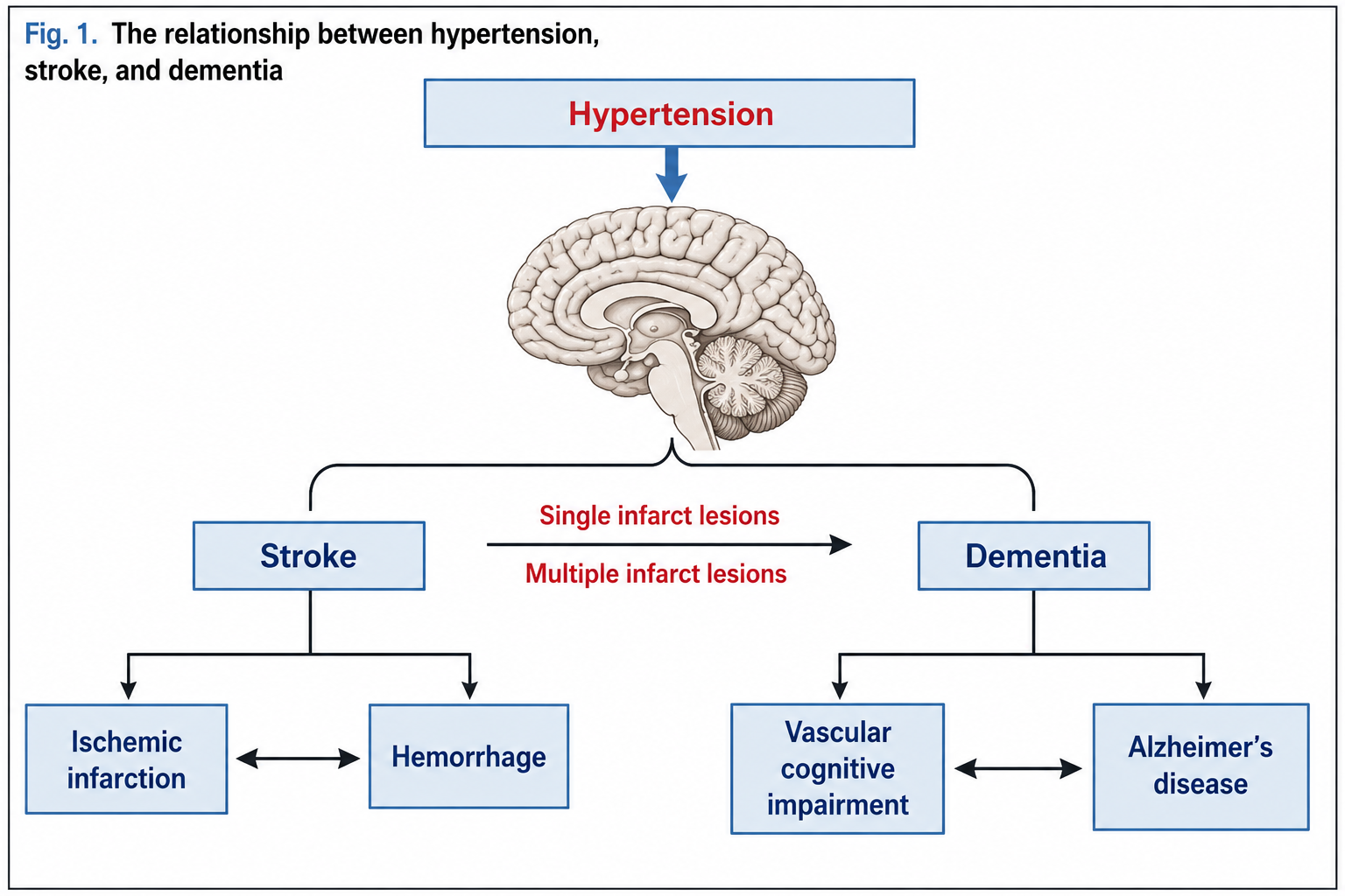
In this article, the relationship between hypertension and cerebrovascular dysfunction is briefly presented, as well as one medication that helps protect the brain from the negative effects of hypertension – vinpocetine.
Cerebral Blood Flow and Adaptive Mechanisms
Branching off from the circle of Willis, larger arteries gradually divide into smaller ones and capillaries that supply all structures of the brain. Like in any other tissues and organs of the body, the blood vessels of the brain are lined with endothelial cells. Arteries and arterioles have 1 or 2 layers of smooth muscle cells that regulate vessel diameter through contractions. The smallest blood vessels – capillaries – do not have smooth muscle cells. Instead, their outer layer is composed of pericytes. Nerve fibers from the autonomic and sensory ganglia of the brain innervate brain arteries and arterioles. Constrictions of smaller blood vessels are regulated by astrocytes.
The brain has a so-called blood-brain barrier – the small blood vessels (capillaries) are impermeable to most substances circulating in the blood. Blood vessels within the brain and on the brain's surface differ in resistance – vessels supplying superficial brain structures (cortex) are almost 3 times more sensitive to various blood flow changes than intracerebral ones.
The brain has practically no energy reserves, so it can function normally only when well supplied with blood and the oxygen and energy-providing substances it contains. There are several adaptive mechanisms that help ensure normal brain function even when various circulatory disorders occur (excluding critical ones). These are:
● functional hyperemia. The intensity of blood flow in various areas of the brain varies. When a specific area of the brain becomes more active, blood flow in that area increases. This phenomenon is called functional hyperemia (4). Studies have shown that neurons, astrocytes, and cells in the blood vessels release various vasodilating substances when specific brain functions are activated, such as nitric oxide, carbon monoxide, prostanoids, cytochrome P450 metabolites, adenosine, and K+ ions (4). In order for the intraparenchymal and meningeal blood vessels of the brain to dilate coordinately, there must be a harmonious interaction between the vascular cells and the adjacent astrocytes (4);
● cerebrovascular autoregulation. Various processes of cerebrovascular autoregulation ensure that cerebral blood flow remains unchanged when the mean arterial blood pressure varies from 60 to 150 mm Hg (7). As known, arterial blood pressure can vary significantly throughout the day (8). In order to maintain optimal blood flow in the brain, intracerebral arterioles constrict when systemic arterial blood pressure increases and dilate when arterial blood pressure decreases. Cerebrovascular autoregulation is ensured only when the myocytes in the vessel walls function normally (myogenic response to changes in blood flow) (9). Under optimal conditions, an increase in intravascular pressure leads to depolarization of myocyte membranes, Ca2+ ions enter the cell, and myocyte contraction is stimulated. In this process, in addition to Ca2+ ions, protein kinase C and Rho kinase also play a role (9, 10);
● endothelial regulation. Endothelial cells of the brain's blood vessels secrete many substances that regulate vascular tone. Nitric oxide, prostacyclin, bradykinin, among others, have vasodilatory effects, while endothelin, an endothelium-derived vasoconstrictor, among others, causes vasoconstriction (11). Vasoactive substances secreted by endothelial cells mainly affect blood vessels at rest (under optimal systemic blood pressure and normal brain activity), but have little effect on ensuring normal brain blood flow in the presence of noticeable fluctuations in systemic blood pressure (11).
Effects of Hypertension on Brain Blood Vessels
Hypertension can negatively affect brain blood vessels in various ways. The main ones are:
● atherosclerosis and lipohyalinosis. Studies have shown that hypertension promotes the formation of atherosclerotic plaques in the brain's arteries and arterioles. For this reason, blood vessels can become blocked, leading to ischemic damage (infarction) (2, 12). There is evidence that hypertension causes fibrinoid necrosis (lipohyalinosis) of penetrating arteries and arterioles in the brain's white matter, associated with small infarctions (lacunes) or hemorrhages (12);
● hypertrophy, remodeling, and stiffening. With persistent hypertension, both systemic and brain arteries undergo changes – the cells in the vessel walls hypertrophy and/or hyperplasia, resulting in indentations of the smooth muscle layer into the vessel lumen. As a result, the vessel walls stiffen, and their diameter decreases (13). Hypertension can also lead to eutrophic remodeling, where the vessel diameter decreases due to rearrangement of smooth muscle cells, without significant changes in their number and size. With prolonged or persistent hypertension, collagen accumulates in the vessel walls, leading to vessel rigidity and stiffening.
Hypertrophy of brain blood vessel walls develops due to increased sympathetic perivascular innervation (12) and because the mechanical increase in intravascular pressure promotes the release of growth factors and molecules supporting oxidative stress and inhibits NO production (14, 15). NO has an antiproliferative effect, so a decrease in its quantity in the vessel walls accelerates myocyte hypertrophy.
One of the main factors related to brain vascular remodeling is angiotensin II. Studies on laboratory animals diagnosed with primary hypertension have shown that administering angiotensin II receptor blockers slows down the intracerebral vascular remodeling processes. A clear positive effect of angiotensin II receptor blockers on myocyte hypertrophy has not been determined. Active free oxygen radicals promote vascular remodeling by increasing the activity of the angiotensin II system, activating myocyte proliferation, and stimulating structural changes in the extracellular matrix.
Hypertrophy and remodeling of brain vascular walls are primarily adaptive measures to maintain optimal cerebral blood flow with changing systemic arterial blood pressure. When these mechanisms are disrupted, cerebral blood flow changes, intracerebral edema develops, and other vascular disorders occur. Prolonged hypertrophy and remodeling processes due to constantly elevated systemic blood pressure become harmful, leading to irreversible changes in the structure of intracerebral vessel walls, reduced lumen diameter, compromised blood flow in certain brain regions, progression of cognitive function impairment, and the potential development of cerebral infarction.
Hypertension negatively affects cerebral blood flow at rest and disrupts adaptive mechanisms. Several studies have found that cerebral blood flow in individuals with hypertension at rest is slower than in individuals with normal blood pressure. The slowing of blood flow can occur due to various reasons, such as aging, decreased cerebral activity (especially in certain areas), vascular constriction, and constant high vascular tone (due to endothelial dysfunction). Slowed blood flow at rest can lead to lacunar infarctions and various central nervous system disorders.
In chronic hypertension, functional hyperemia is impaired. When the activity of a specific brain region intensifies, blood flow to that region does not increase. This impairment can lead to the progression of various cognitive function impairments.
Due to constantly elevated or frequently increased systemic blood pressure, dysfunction of cerebrovascular endothelium develops, disrupting endothelium-dependent vasodilation of cerebral blood vessels. Patients with hypertension also experience impaired autoregulatory mechanisms of cerebral blood vessels. Adequate blood supply to normal cerebral structures requires higher perfusion pressure. Therefore, when systemic blood pressure decreases (even to a normal level), cerebral blood flow becomes insufficient, leading to ischemia. The most sensitive areas to this type of ischemia are the white matter regions adjacent to the cerebral ventricles, as this is where the arteries descending from the cerebral meninges and ascending from the basal ganglia branch out. Studies have shown that the extent of periventricular white matter damage in hypertensive individuals correlates with the degree of autoregulatory dysfunction and cognitive function impairment.
The Role of Oxidative Stress in the Pathophysiology of Hypertension
In recent years, numerous studies have provided evidence that oxidative stress plays a significant role in various pathophysiological processes related to hypertension. It has been found that markers of oxidative stress increase in the blood in primary arterial hypertension, renovascular hypertension, secondary hypertension, and preeclampsia. Experimental studies on hypertensive laboratory mice have shown that endothelial dysfunction in cerebral blood vessels partially develops due to the action of free oxygen radicals. The main source of these radicals in the brain is the NADPH oxidase enzymatic system. Mitochondrial enzymes and xanthine oxidase also produce some of these radicals.
Studies have revealed that damage to cerebral blood vessels occurs when free oxygen radicals combine with NO, forming peroxynitrites that cause DNA damage, lipid peroxidation, and disrupt intracellular and extracellular protein functions of the vessel wall. The latest research data have revealed that peroxynitrites play a significant role in cerebrovascular circulation disorders related to the angiotensin II system.
The Relationship Between Hypertension and Alzheimer's Disease
It is commonly believed that Alzheimer's disease is a neurodegenerative pathology that develops when normal neuron activity is disrupted due to the accumulation of beta-amyloid in the brain and changes in the neuronal cytoskeleton, resulting in progressive cognitive impairment (3). The latest research data have revealed that one of the factors that promotes the development of Alzheimer's disease is chronic hypertension, especially when it occurs in middle-aged individuals.
Neurovisual studies have shown that individuals with chronic hypertension have more areas of atrophy, amyloid plaques, and changes in the neuronal cytoskeleton in the brain (32). It has been established that in cases where the brain of a person with Alzheimer's disease shows changes typical of hypertension, the cognitive function deficit is greater than in individuals without high blood pressure (33). It has also been observed that individuals with Alzheimer's disease more frequently have ischemic lesions and more advanced vascular atherosclerosis in their brains (34). The exact relationship between Alzheimer's disease and hypertension is not clear and should be elucidated through further studies.
How to Protect the Brain from the Negative Effects of Hypertension? Benefits of Vinpocetine
To protect the brain from the negative effects of hypertension, it is essential to adequately control blood pressure and take all possible preventive measures against hypertension. Unfortunately, this is not always possible. Persistent hypertension causes various structural changes in the brain, deteriorating cognitive functions. In such cases, medications should be prescribed to help protect the brain from the adverse effects of hypertension and reduce cognitive impairments. One such medication is vinpocetine.
Vinpocetine (ethyl apovincaminate) is a drug that selectively stimulates cerebral blood flow, oxygen supply, improves blood rheological properties, reduces platelet aggregation, inhibits the enzyme phosphodiesterase, and has antithrombotic effects. Studies involving patients with various degrees of vascular dementia have shown that vinpocetine significantly improved cognitive functions, memory, speech, and reading (35, 36). A study conducted in Japan found that administering vinpocetine to patients who had suffered a stroke led to faster recovery of the damaged central nervous system functions (including cognitive functions) (37). This drug is also effective for rare forms of dementia. One study included individuals with the rare Binswanger's type of dementia (characterized by small infarcts in the white matter of the brain). It was found that administering vinpocetine significantly improved blood flow and oxygen supply to structurally altered brain areas, reducing cognitive deficits (38).
Vinpocetine is a potent vasodilator that directly relaxes smooth muscle in blood vessels. In cerebrovascular pathology, vinpocetine helps improve blood flow in the brain. This drug is also beneficial for patients at risk of thrombus formation, as it reduces platelet and red blood cell aggregation and increases red blood cell membrane flexibility (39).
Studies have shown that vinpocetine has an antihypoxic effect. Unlike other similar drugs, vinpocetine does not cause the "steal" phenomenon (i.e., a condition where the ischemic area receives more blood supply at the expense of other brain regions). Animal models have shown that in cases of anoxia, vinpocetine helps reduce brain edema and increase survival rates (40).
The usual dose of vinpocetine is 15–45 mg/day. Visual studies have shown that when this drug is taken orally, it reaches the brain within 10 minutes and distributes heterogeneously, meaning more in areas with the most impaired blood flow (40). The drug is generally well tolerated. Rare side effects include skin rashes, itching, and gastrointestinal symptoms. These reactions are usually not intense, and patients do not discontinue the medication. It is important to note that vinpocetine does not cause tolerance, meaning its effects do not diminish with long-term use. It should be noted that the maximum therapeutic effect is not immediate but occurs after several weeks of treatment, so the drug should be taken for a long time.
Conclusion
Hypertension is one of the main health problems in many developed countries. When prescribing treatment, it is essential to consider not only adequate blood pressure reduction but also measures to reduce the consequences of hypertension in the body. One drug that should be included in the treatment plan for a patient with hypertension is vinpocetine. This plant-based medication improves blood circulation in the brain, supplies it with oxygen and other energy substances, effectively reducing cognitive function disorders caused by circulatory disorders.
Prepared by Dr. Berta Zaleckienė
Source: "Internistas"
