
Acute Leukemia
Acute leukemia is rarely encountered in clinical practice. Because this oncological disease progresses rapidly, over weeks or even days, early diagnosis is particularly important. If diagnosis is delayed, the complications of the disease may, in certain cases, become difficult to manage and life-threatening. If acute leukemia is suspected, the patient should be referred to a hematologist as soon as possible for further evaluation and treatment.
Patients receiving palliative treatment should be closely monitored by their family physician when they are not hospitalized for clinical signs of anemia, possible bleeding, or infectious complications. If symptoms develop, patients should be promptly referred for blood transfusions or receive appropriate antibiotic therapy. Because the condition of these patients can change rapidly and their treatment and care are highly specific, close cooperation between the family physician or internist caring for the patient and the hematologist is essential.
What Is Acute Leukemia?
Acute leukemia is an oncological blood disease that develops when normal hematopoietic stem cells transform into primitive, undifferentiated cells with an abnormal lifespan—blasts. Blasts replace normal blood cells, resulting in deficiencies of normal blood cells, including anemia, thrombocytopenia, and neutropenia, often accompanied by leukocytosis when the elevated leukocyte count is due to blasts. Blasts may infiltrate various internal organs and tissues, including the liver, spleen, lymph nodes, central nervous system (CNS), kidneys, and others, leading to dysfunction of these organs. Acute leukemia may arise from the myeloid lineage, as acute myeloid leukemia (AML), or from the lymphoid lineage, as acute lymphoblastic leukemia (ALL).
Epidemiology
Acute myeloid leukemia is the most common leukemia in adults. In Europe, its incidence is 5–8 cases per 100,000 population per year. The risk of developing AML increases with age, with the highest incidence among people older than 70 years, reaching 15–25 cases per 100,000 population per year. Mortality is 4–6 cases per 100,000 population per year. In children, AML accounts for approximately 10–15% of all cases.
Acute lymphoblastic leukemia is the most common leukemia in children aged 3–7 years. It accounts for 20% of all acute leukemias in adults. There are two peaks in incidence: around the age of 40 and among people older than 80 years. The incidence of ALL is 0.7–1.8 cases per 100,000 population per year.
Etiology and Risk Factors
The exact causes contributing to the development of these diseases remain unclear. Factors predisposing to leukemia include ionizing radiation and genetic disorders, including Down, Klinefelter, Fanconi, and Bloom syndromes. Air pollution, viruses, smoking, and chemicals may also contribute to the development of acute leukemia.
Acute myeloid leukemia may be primary, developing de novo, or secondary, developing in patients with other hematological diseases, such as myelodysplasia or other myeloproliferative disorders, or following chemotherapy. Primary and secondary AML differ in their genetic markers and prognosis. Secondary leukemia has a poorer prognosis.
Clinical Presentation
Symptoms may develop over a period ranging from several days to several weeks before diagnosis. The most common symptoms result from impaired hematopoiesis or tumor-related intoxication.
- Syndrome of normal hematopoietic insufficiency. This develops because of inadequate production of normal blood cells. Clinical symptoms of anemia occur. In the presence of thrombocytopenia, bleeding may manifest as petechiae, bruising of the skin, epistaxis, bleeding from the gums, or irregular and heavy menstrual bleeding. Gastrointestinal bleeding and hematuria are uncommon. Because these patients are immunosuppressed as a result of the disease itself and the inadequate production of normal leukocytes, infectious complications, including bacterial and fungal infections, are common and may progress rapidly and become life-threatening.
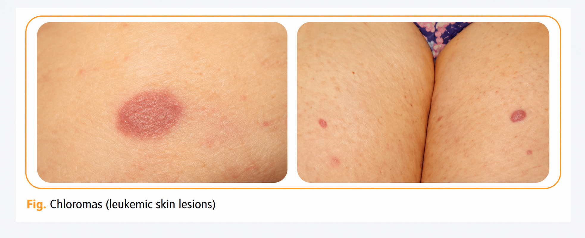
- Tumor infiltration syndrome. This develops as a result of blast infiltration into various organs and tissues. CNS involvement (neuroleukemia) or leukemic meningitis is rare but may occur, presenting with headache, vomiting, seizures, and other symptoms. Infiltration of the bone marrow and periosteum may cause bone or joint pain, particularly in children with ALL. Extramedullary blast infiltration may lead to splenomegaly, hepatomegaly, and leukemic skin involvement, presenting as a nonpruritic skin rash—chloromas. Gingival hyperplasia frequently develops in AML.
- Tumor intoxication syndrome. The symptoms are nonspecific and may include fatigue, general weakness, low-grade fever, weight loss, and increased sweating. They result from hypermetabolic reactions occurring in the body.
- Leukostasis syndrome. Symptoms develop because of a large number of blasts circulating in the peripheral blood, most commonly when the leukocyte count exceeds 100 × 10⁹/L. Because of their large number, the cells aggregate and form microthrombi, which may impair blood flow in the CNS, lungs, genital organs, and eyes.
Automated Peripheral Blood Test in a Patient With Acute Leukemia
(LSMU MA Laboratory Medicine Clinic, Hematology and General Cytology Laboratory)
| Analyte | Result | Unit | Reference Range |
|---|---|---|---|
| RBC | 2.55 | ×10¹²/L | 3.9–5.1 |
| HGB | 72 | g/L | 119–146 |
| HCT | 21 | % | 37–44 |
| MCV | 82.4 | fL | 83–98 |
| MCH | 28.2 | pg | 27–32.3 |
| MCHC | 343 | g/L | 318–347 |
| RDW-CV | 15.2 | % | 12.1–14.3 |
| WBC | 2.37 | ×10⁹/L | 4.4–9.7 |
| NE% | 21.1 | % | 43–74 |
| LY% | 31.2 | % | 18–46 |
| MO% | 43.5 | % | 4.2–11.8 |
| EO% | 2.1 | % | 0.2–5.3 |
| BA% | 2.1 | % | 0.1–1 |
| NE# | 0.5 | ×10⁹/L | 2–7.2 |
| LY# | 0.74 | ×10⁹/L | 1.3–3.4 |
| MO# | 1.03 | ×10⁹/L | 0.3–0.8 |
| EO# | 0.05 | ×10⁹/L | 0.01–0.4 |
| BA# | 0.05 | ×10⁹/L | 0.01–0.07 |
| IG% | 1.7 | % | 0–0.6 |
| IG# | 0.04 | ×10⁹/L | 0–0.06 |
| NRBC% | 0 | /100 leukocytes | 0–0 |
| NRBC# | 0 | ×10⁹/L | 0–0 |
| PLT | 21 | ×10⁹/L | 173–390 |
| PCT | – | % | 0.18–0.39 |
| MPV | – | fL | 9.1–11.9 |
| PDW | – | fL | 9–15.4 |
| P-LCR | – | % | 17.5–42.3 |
Peripheral Blood Cytomorphological Examination
(Modified Giemsa Method)
| Analyte | Result | Unit | Reference Range |
|---|---|---|---|
| Blasts | 40 | % | – |
| Metamyelocytes | 2 | % | – |
| Band neutrophils | 1 | % | 1–5 |
| Segmented neutrophils | 21 | % | 40–70 |
| Eosinophils | 1 | % | 0–5 |
| Basophils | 0 | % | – |
| Monocytes | 17 | % | 3–12 |
| Lymphocytes | 18 | % | 20–45 |
Diagnosis
A complete blood count (CBC) may raise suspicion of the disease: leukocytosis, a low platelet count, and anemia or pancytopenia may be detected. If a differential leukocyte count can be performed, the presence of blasts is a diagnostic marker. Other specific tests performed by a hematologist help determine the type of leukemia and the prognosis of the disease.
- Complete blood count and peripheral blood smear. This is the first test that may help raise suspicion of acute leukemia. Pancytopenia may be observed on the CBC, while blasts may be detected in the leukocyte differential. Blasts may account for up to 90% of all leukocytes. At the onset of the disease, blasts may be absent from the peripheral blood. Other causes of pancytopenia should also be considered, including viral infections such as infectious mononucleosis, vitamin B12 and folic acid deficiency, and others. Very rarely, infectious diseases such as tuberculosis may cause a leukemoid reaction on the CBC, with blasts in the peripheral blood.
- Bone marrow aspiration and biopsy. Leukemia is diagnosed when blasts account for >20%. These tests are used to determine the type of leukemia.
- Histochemical studies, immunophenotyping, and genetic mutations. Depending on the type of acute leukemia, blasts differ in their morphology, immunophenotype, and cytogenetic characteristics. Specific monoclonal antibodies present on B- and T-lineage cells are examined. Flow cytometric immunophenotyping distinguishes the type of leukemia as acute myeloid or lymphoid leukemia. It also differentiates individual types of acute myeloid leukemia (AML), which differ in their clinical course and treatment. Cytogenetic studies are essential for all patients with acute leukemia because they help determine prognosis and guide treatment.
Automated Peripheral Blood Test in a Patient With Acute Leukemia
(LSMU MA Laboratory Medicine Clinic, Hematology and General Cytology Laboratory)
| Analyte | Result | Unit | Reference Range |
|---|---|---|---|
| RBC | 2.18 | ×10¹²/L | 3.9–5.1 |
| HGB | 73 | g/L | 119–146 |
| HCT | 22 | % | 37–44 |
| MCV | 100.9 | fL | 83–98 |
| MCH | 33.5 | pg | 27–32.3 |
| MCHC | 332 | g/L | 318–347 |
| RDW-CV | 15.5 | % | 12.1–14.3 |
| WBC | 32.54 | ×10⁹/L | 4.4–9.7 |
| NE% | – | % | 43–74 |
| LY% | – | % | 18–46 |
| MO% | – | % | 4.2–11.8 |
| EO% | – | % | 0.2–5.3 |
| BA% | 0.1 | % | 0.1–1 |
| NE# | – (calculated by cytomorphological examination) | ×10⁹/L | 2–7.2 |
| LY# | – (calculated by cytomorphological examination) | ×10⁹/L | 1.3–3.4 |
| MO# | – | ×10⁹/L | 0.3–0.8 |
| EO# | – | ×10⁹/L | 0.01–0.4 |
| BA# | 0.03 | ×10⁹/L | 0.01–0.07 |
| IG% | – | % | 0–0.6 |
| IG# | – | ×10⁹/L | 0–0.06 |
| NRBC% | 1.1 | /100 leukocytes | 0–0 |
| NRBC# | 0.37 | ×10⁹/L | 0–0 |
| PLT | 272 | ×10⁹/L | 173–390 |
| PCT | 0.29 | % | 0.18–0.39 |
| MPV | 10.7 | fL | 9.1–11.9 |
| PDW | 12.5 | fL | 9–15.4 |
| P-LCR | 29.8 | % | 17.5–42.3 |
Peripheral Blood Cytomorphological Examination
(Modified Giemsa Method)
| Analyte | Result | Unit | Reference Range |
|---|---|---|---|
| Blasts | 85 | % | – |
| Band neutrophils | 0 | % | 1–5 |
| Segmented neutrophils | 5 | % | 40–70 |
| Eosinophils | 0 | % | 0–5 |
| Basophils | 0 | % | – |
| Monocytes | 2 | % | 3–12 |
| Lymphocytes | 8 | % | 20–45 |
Prognostic Factors
After AML is diagnosed, the patient’s age and cytogenetic and molecular changes are the most important prognostic factors determining the course of the disease.
Treatment
The main goal of treatment is to destroy malignant cells and achieve disease remission and a complete hematological response. Hematological remission is achieved when, after treatment, <5% blasts remain in the bone marrow and blood counts recover, with platelets >100 × 10⁹/L, neutrophils >1.5 × 10⁹/L, and Hgb >100 g/L. When complete hematological remission lasts longer than 5 years, the patient is considered cured.
Treatment outcomes are poorer in patients older than 65–70 years. This is because, at this age, the disease is often more resistant to treatment and intensive chemotherapy is also poorly tolerated. If a patient has comorbidities or is in poor physical condition, achieving remission may not be the treatment goal, and symptomatic palliative treatment is used instead. Treatment may consist of a single chemotherapeutic agent (low-dose cytarabine, hydroxycarbamide) or only blood component transfusions and antibiotic therapy.
AML and ALL are treated with chemotherapy and supportive (symptomatic) treatment. Specific chemotherapy for ALL consists of four treatment phases: remission induction, CNS prophylaxis, consolidation, and maintenance treatment.
Specific Treatment
- Chemotherapy. Cytotoxic drugs are not selective for malignant cells and exert similar toxic effects on all hematopoietic cells, causing severe bone marrow suppression and resulting in profound, prolonged pancytopenia and associated complications.
- Allogeneic hematopoietic stem cell transplantation (HSCT). This is used in patients younger than 65 years who belong to intermediate- and high-risk groups and have achieved remission. Post-transplant mortality is approximately 20–25%.
- Autologous HSCT. This is used in patients younger than 65 years who belong to intermediate- and high-risk groups and have achieved remission. Mortality after autologous HSCT is much lower than after allogeneic HSCT, but the relapse rate is significantly higher, reaching up to 50%.
Supportive Treatment
- Blood component transfusions. Prophylactic platelet transfusions are administered when the platelet count is <10 × 10⁹/L. In the presence of hemorrhagic syndrome or infectious complications, platelet transfusion is administered when the platelet count is <20 × 10⁹/L. Red blood cell transfusions are administered when Hgb is <70 g/L or <80 g/L in the presence of signs of tissue hypoxia. Red blood cell transfusions should be avoided while the patient’s leukocyte count is >100 × 10⁹/L because of the potential risk of hyperviscosity and thrombus formation.
- Antibiotic therapy and antifungal medications. Patients frequently have profound neutropenia and immunosuppression. Immunosuppression develops because of neutropenia, hypogammaglobulinemia, impaired immune cell function, the disease itself, and its treatment. Infections in these patients progress rapidly, without a clinical prodrome, and are a major cause of death among patients with acute leukemia. During periods of neutropenia, patients are advised to avoid raw vegetables and fruits. Visits to these patients are restricted, disposable masks are recommended, and good personal hygiene of both patients and their caregivers is particularly important. Protocols for the prevention of bacterial and fungal infections vary among institutions depending on the predominant pathogens. In febrile neutropenia, defined as a temperature >38°C lasting longer than 1 hour and neutropenia <0.5 × 10⁹/L, treatment with intravenous antibiotics must be initiated within one hour, after obtaining blood and urine cultures and cultures from other sites that may represent a source of infection. Febrile neutropenia is a life-threatening condition, and with every hour of delayed treatment, the patient’s risk of death increases. Treatment is initiated with broad-spectrum antibiotics active against gram-negative and gram-positive microorganisms (ceftazidime, imipenem). If necessary, the spectrum of antibiotic therapy is broadened and subsequently adjusted according to culture results. If antibacterial treatment is ineffective, empirical antifungal treatment is initiated. Confirmed aspergillosis is treated with amphotericin, voriconazole, or caspofungin. Candidiasis is treated with fluconazole, itraconazole, or posaconazole.
- Tumor lysis syndrome. The syndrome may develop after chemotherapy is initiated because of the rapid destruction of leukemic cells. During the syndrome, plasma uric acid concentration increases, and hyperphosphatemia, hyperkalemia, and hypocalcemia may occur. These abnormalities may lead to acute renal failure. To prevent this, adequate hydration, allopurinol, or rasburicase is administered.
- Psychological support for the patient and family members helps them cope with the diagnosis and the difficulties associated with treatment.
Prognosis
Cure is possible in both types of leukemia, particularly in younger patients. The prognosis is poorer in infants, older patients (>60 years), and those with comorbid conditions such as liver involvement, renal failure, CNS or testicular involvement, myelodysplasia, or a high leukocyte count (>25 × 10⁹/L) at diagnosis.
The expected survival of untreated patients is 3–6 months. Survival prognosis depends on various factors, including the patient’s age, karyotype, specific cytogenetic and molecular changes, response to treatment, and physical condition. Approximately half of patients older than 60 years achieve remission, but the disease relapses in 80–90% of cases.
Practical Advice for Physicians
- If a complete blood count reveals pancytopenia with blasts or blasts alone, acute leukemia should be suspected and the patient urgently referred to a hematologist.
- Patients monitored on an outpatient basis should be followed for clinical symptoms of anemia, hemorrhagic syndrome, and infections.
- Platelet transfusions should be administered when the platelet count is <10 × 10⁹/L. In the presence of hemorrhagic syndrome or infectious complications, they should be administered when the platelet count is <20 × 10⁹/L.
- Red blood cell transfusions are administered when Hgb is <70 g/L or <80 g/L in the presence of clinical signs of anemia.
- Febrile neutropenia (FN) is defined as a temperature >38°C lasting longer than 1 hour and neutropenia <0.5 × 10⁹/L.
- Febrile neutropenia is a life-threatening condition; therefore, the patient should be referred to a tertiary-level hospital for treatment as quickly as possible.
- Psychological support for patients and their relatives is a very important aspect of helping them cope with the disease and its consequences. Referral to appropriate specialists and participation in the oncohematological patient association “Kraujas” are recommended.
Source: Lietuvos gydytojo žurnalas
Eglė Simaškienė, LSMU MA Department of Hematology
