
Acute Viral Encephalitis
Prepared according to Tyler KL. Acute Viral Encephalitis. N Engl J Med. 2018;379(6):557–566.
Dr. Mindaugas Petrašiūnas
Introduction
Encephalitis is an infectious or inflammatory disorder of the brain parenchyma that typically presents with fever, headache, altered consciousness, changes in mental status, and other symptoms. This syndrome can be caused by many factors, one of the most common being neurotropic viruses (1). This review describes the main principles of the diagnosis and treatment of acute viral encephalitis.
Epidemiology
The incidence of acute viral encephalitis varies across different regions of the world and is approximately 7 cases per 100,000 population. It is believed that these figures do not reflect the true incidence and clinical diversity of the disease because of insufficient diagnosis and reporting of cases. The cause of approximately 50% of all cases of encephalitis remains unclear (4).
Neurotropic viruses are responsible for 20–50% of encephalitis cases in which the cause has been identified. Herpes simplex virus (HSV) causes 50–75% of encephalitis cases with an identified viral etiology, while varicella-zoster virus (VZV), enteroviruses, and arboviruses account for the majority of the remaining cases of viral encephalitis (5).
HSV encephalitis affects patients of all ages, does not follow a typical seasonal pattern, and is not associated with a specific geographical region. Arbovirus-induced encephalitis, including tick-borne encephalitis virus (TBEV) infection, shows annual variation in incidence, occurs seasonally, and has a prevalence that depends on the geographical region. TBEV-induced encephalitis is one of the most common forms of flavivirus-induced encephalitis worldwide (6).
Lithuania is an endemic region for tick-borne diseases, with approximately 300–600 cases of tick-borne encephalitis registered annually. In 2016, the incidence of tick-borne encephalitis alone was 22.1 cases per 100,000 population and reached 32.3 cases per 100,000 among rural residents (7). Therefore, it is important to remember that from April to December, tick-borne encephalitis is one of the most common causes of central nervous system (CNS) infections in Lithuania, with approximately 90% of all tick-borne encephalitis cases in Lithuania occurring between June and October (7).
Meningitis and Encephalitis
The most important distinguishing feature between meningitis and encephalitis is the presence of impaired brain function. Patients with meningitis may experience headache and lethargy, but their cerebral functions remain intact. In encephalitis, cerebral functions are affected, resulting in changes in mental status, behavior, personality, speech, motor function, and other manifestations.
Seizures occur only in meningitis and should not be considered a sign of encephalitis alone. Unfortunately, the differential diagnosis of meningitis and encephalitis syndromes in clinical practice is often challenging because parenchymal and meningeal inflammation can present with clinical features of both processes. A patient with features of both syndromes is usually described according to the predominant clinical presentation, or the term "meningoencephalitis" is used to encompass both syndromes.
The differential diagnosis of aseptic meningitis and encephalitis is important because certain viral agents are characteristic of meningitis and encephalitis. For example, enteroviruses, VZV, HSV-2, influenza, and measles viruses are more common in aseptic meningitis (8).
Clinical Presentation of Viral Encephalitis
Clinical case descriptions indicate that the clinical presentation of HSV-induced encephalitis does not differ significantly from that of encephalitis of other etiologies. However, HSV encephalitis has been associated with greater cerebrospinal fluid pleocytosis and focal abnormalities on electroencephalography (EEG) and neuroimaging studies (9).
A review of adult patients with encephalitis who had temporal lobe abnormalities on MRI revealed that HSV encephalitis was more likely to be associated with the following features: older age, acute clinical presentation (88% of HSV encephalitis cases compared with 64% of encephalitis cases caused by other viruses), fever (80% and 49%, respectively), gastrointestinal symptoms (37% and 19%, respectively), and less frequent ataxia (18% and 33%, respectively) and rash (2% and 15%, respectively).
The data also showed that male patients were more likely to have HSV rather than autoimmune encephalitis (50% and 14%, respectively) and were less likely to present with psychosis (5% and 20%, respectively) or rash (2% and 21%, respectively). Most neurological symptoms, including altered consciousness, aphasia, hallucinations, and motor disturbances, did not differ significantly among patients with encephalitis of different etiologies. There were no significant differences in MRI findings between different etiologies of encephalitis, although patients with non-HSV encephalitis more frequently had bilateral hippocampal lesions and lesions extending beyond the hippocampus (10).
It has been noted that certain viruses causing encephalitis tend to produce regional MRI abnormalities and may be suspected based on the patterns of these changes. Based on retrospective MRI findings in patients with encephalitis, focal and generalized disease profiles have been identified. The focal profile includes features and symptoms attributable to specific brain regions, whereas the generalized profile describes diffuse cerebral dysfunction, including diffuse cerebral edema, generalized seizures, and psychosis. Some authors suggest that this classification method may help select diagnostic tests for specific viruses or facilitate more rapid diagnosis of non-viral causes (10–12).
Diagnosis
Cerebrospinal fluid (CSF) examination is the initial diagnostic step in patients with suspected viral encephalitis. Evaluation of CSF pressure and its physical characteristics can provide clues regarding the possible etiology. In viral encephalitis, CSF pressure is usually slightly elevated, while the fluid appears clear or slightly cloudy.
Laboratory examination of the CSF in viral encephalitis reveals cytosis (approximately 50–500 × 10⁶/L, with a predominance of lymphocytes), normal or slightly elevated glucose levels, and moderately increased protein concentrations. Following CSF analysis, if viral encephalitis is suspected, polymerase chain reaction (PCR) testing should be performed to detect HSV-1, HSV-2, and enteroviruses. Other tests, including serological tests, for example for arboviruses (EEE), are performed according to the clinical presentation, season, and geographical region (1, 8).
PCR is used to detect DNA and RNA viruses. PCR testing of CSF is commonly performed to detect HSV-1, HSV-2, VZV, enteroviruses, and, in young children under 3 years of age, human parechoviruses. If these tests do not establish the etiology of encephalitis, additional PCR testing of CSF may be indicated to detect cytomegalovirus (CMV), human herpesvirus (HHV)-6, HHV-7, Epstein-Barr virus (EBV), and HIV. These tests should be performed immediately in immunosuppressed patients.
When specific pathogens are suspected, PCR testing may be performed using specific samples: throat or nasopharyngeal samples for adenovirus, influenza, and measles infections; saliva for epidemic parotitis or rabies infections; and stool samples for the detection of enteroviral infections.
Serological tests, including serum and CSF examinations performed during the acute and convalescent stages, are crucial for diagnosing arboviruses and other specific viruses, such as VZV, EBV, measles, epidemic parotitis, rubella, and rabies. In cases of tick-borne encephalitis, the diagnosis is usually established through serological testing. When CNS involvement occurs, EEV IgM antibodies are detected in approximately 90% of patients' blood serum samples and in 50% of CSF samples (1, 7).
Radiological abnormalities are not always detected on neuroimaging studies, including computed tomography (CT) and magnetic resonance imaging (MRI), in patients with encephalitis. CT is useful for excluding intracranial processes, whereas MRI is used to identify demyelinating processes that may present with similar changes in mental status, such as progressive multifocal leukoencephalopathy. When abnormalities are detected on neuroimaging, their characteristics may provide clues to specific etiologies:
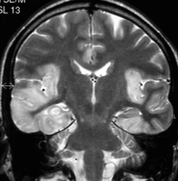
• Temporal lobe involvement is characteristic of HSV-1 encephalitis, although similar abnormalities may be caused by other herpesviruses, including VZV, EBV, or HHV-6 (Figure 1);
• Involvement of the thalamus and basal nuclei in encephalitis may be caused by respiratory viral infections, Creutzfeldt-Jakob disease, arboviral infections, or tuberculosis;
• Hydrocephalus may indicate a non-viral etiology, including bacterial, fungal, or parasitic causes;
• Multifocal supratentorial white matter abnormalities on MRI are characteristic of post-infectious encephalitis.
Electroencephalography (EEG) findings are often abnormal in patients with acute encephalitis. Focal abnormalities in the temporal regions suggest a probable HSV-1 etiology (8).
Figure 1. Asymmetric T2 hyperintensity in the temporal lobes of a patient with HSV-1 encephalitis (26).
 Treatment Strategy
Patients with encephalitis should be closely monitored to ensure adequate oxygenation, airway patency, and circulatory support, as well as appropriate management of fever, arrhythmias, cerebral edema, increased intracranial pressure, and focal and generalized seizures.
Several empirical and specific treatment guidelines are currently available for patients with viral encephalitis (4, 13, 14). Unfortunately, only a few existing therapies have been investigated in randomized controlled clinical trials. For example, in the guidelines of the Infectious Diseases Society of America (4), only the use of acyclovir is classified as a Level A recommendation (good evidence to support the recommendation) with Level I quality of evidence (evidence obtained from one or more randomized controlled trials). Recommendations from other organizations are similar (13, 14).
The Level A recommendations in the Infectious Diseases Society of America guidelines include the empirical use of acyclovir in all patients with suspected encephalitis. The British national guidelines for the treatment of viral encephalitis also recommend empirical acyclovir therapy but, like the Infectious Diseases Society of America guidelines, acknowledge that this recommendation is based on lower-quality evidence than data obtained from randomized controlled trials (14).
Ganciclovir or foscarnet is recommended in some guidelines for encephalitis associated with CMV and HHV-6, and acyclovir is recommended for VZV encephalitis. However, these recommendations are based on moderate-quality evidence from expert opinion and descriptive studies (4, 14). Due to the lack of high-quality evidence, guidelines from other associations do not provide specific treatment recommendations for CMV-, HHV-6-, and VZV-associated encephalitis (13).
Previously, the initial intravenous acyclovir regimen for adult patients with HSV encephalitis was 10 mg/kg every 8 hours for 10 days in patients with normal kidney function. However, because of the high risk of recurrence, the duration of acyclovir therapy was initially extended from 10 to 14 days, and it is currently recommended for up to 21 days. Neither increasing the acyclovir dose to 15 mg/kg every 8 hours in adults nor long-term therapy with valacyclovir (2 mg every 8 hours for 90 days) improves outcomes in adults.
However, in children aged 3 months to 12 years diagnosed with HSV encephalitis, a higher dose of acyclovir (20 mg/kg every 8 hours for 21 days) results in better outcomes and fewer recurrences than lower doses.
Prevention Tactics
Due to the lack of effective, evidence-based treatment models for most neurotropic viral infections, prevention of these diseases is particularly important. Effective vaccines are currently available against many neurotropic viruses, including poliovirus, rabies, measles, mumps, epidemic parotitis, rubella, influenza, VZV, and several neurotropic flaviviruses, including EEV, which is particularly relevant in Lithuania.
Vaccines against West Nile, dengue, and Zika viruses are currently being evaluated in clinical trials. There are data demonstrating the effectiveness of newer vaccines in reducing the number of encephalitis cases and associated complications. A 5-year vaccination campaign in Nepal aimed at preventing Japanese encephalitis demonstrated a 78% reduction in the number of cases. A universal VZV vaccination program introduced in Germany in 2004 for 1-year-old children resulted in an approximately 60% reduction in neurological complications associated with varicella infection. Rotavirus vaccination in the USA, recommended for infants, resulted in a 4% lower rate of seizure-related hospitalizations among children under 5 years of age, compared with 16% before vaccination.
Outcomes
The outcome of acute viral encephalitis remains unfavorable. Factors associated with an increased risk of an unfavorable outcome include immunosuppressive conditions, a Glasgow Coma Scale score of 8 or lower, admission to the intensive care unit, and age over 65 years.
In HSV-1-induced encephalitis, factors associated with an increased risk of an unfavorable outcome include coma, a delay of >24 hours in initiating acyclovir therapy, and older age. Other findings from MRI, EEG, or cerebrospinal fluid examinations did not have prognostic significance for outcomes.
Unfortunately, despite evidence that early initiation of acyclovir therapy improves outcomes in HSV encephalitis, delays in treatment occur relatively frequently. In a study conducted in Canada, the average time to initiation of acyclovir therapy was 21 hours for all patients with suspected HSV encephalitis and 11 hours for those with confirmed HSV. In a study conducted in the USA, only 29% of patients with suspected encephalitis received acyclovir in the emergency department. A multicenter study conducted in Europe found that treatment was initiated within 48 hours of symptom onset in only 45% of patients with HSV-induced encephalitis.
Factors contributing to delays in medication administration include prolonged waiting times for brain imaging studies, the absence of significant cerebrospinal fluid pleocytosis, and other factors, such as severe concurrent illnesses or alcohol abuse. Inappropriate initial acyclovir dosing is administered to up to 75% of children and 24% of adults treated empirically for suspected viral encephalitis.
Conclusions
Viral encephalitis is a dangerous disease that imposes a significant economic burden. New diagnostic technologies and strategies are being developed to accurately and rapidly identify the causative pathogen and differentiate viral encephalitis from similar diseases. Treatment remains largely empirical, with the exception of acyclovir administration for HSV-induced encephalitis. There is a substantial need for effective, evidence-based prevention and treatment methods to prevent viral encephalitis and its complications.
Publication "Internistas," No. 8, 2018.
References: 1. Tyler KL. Acute Viral Encephalitis. N Engl J Med. 2018;379(6):557-566. 2. Hasbun R, Rosenthal N, Balada-llasat JM, et al. Epidemiology of Meningitis and Encephalitis in the United States, 2011-2014. Clin Infect Dis. 2017;65(3):359-363. 3. Vora NM, Holman RC, Mehal JM, et al. Burden of encephalitis-associated hospitalizations in the United States, 1998-2010. Neurology. 2014; 82:443-451 4. Tunkel AR, Glaser CA, Bloch KC, et al; Infectious Diseases Society of America. The management of encephalitis: clinical practice guidelines by the Infectious Diseases Society of America. Clin Infect Dis. 2008; 47:303-327. 5. George BP, Schneider EB, Venkatesan A. Encephalitis hospitalization rates and inpatient mortality in the United States, 20002010. PLoS One 2014; 9(9):e104169 6. Kunz C, Heinz FX. Tick-born encephalitis. Vaccine. 2003 Apr; 21(1): S1/1–2 7. Center for Communicable Diseases and AIDS. Incidence of communicable diseases in Lithuania in 2016. 2016; 93-98. 8. Gluckman SJ. Viral encephalitis in adults. Post TW, ed. UpToDate. Waltham, MA: UpToDate Inc. 9. Whitley RJ, Soong SJ, Linneman C Jr, Liu C, Pazin G, Alford CA. Herpes simplex encephalitis. JAMA 1982; 247:31720. 10. Chow FC, Glaser CA, Sheriff H, et al. Use of clinical and neuroimaging characteristics to distinguish temporal lobe herpes simplex encephalitis from its mimics. Clin Infect Dis 2015; 60:137783. 11. Glaser CA, Honarmand S, Anderson LJ, et al. Beyond viruses: clinical profiles and etiologies associated with encephalitis. Clin Infect Dis 2006; 43:156577. 12. Beattie GC, Glaser CA, Sheriff H, et al. Encephalitis with thalamic and basal ganglia abnormalities: etiologies, neuroimaging, and potential role of respiratory viruses. Clin Infect Dis 2013; 56:82532. 13. Steiner I, Budka H, Chaudhuri A, et al. Viral meningoencephalitis: a review of diagnostic methods and guidelines for management. Eur J Neurol 2010;17(8):999 1009. 14. Solomon T, Michael BD, Smith PE, et al. Management of suspected viral encephalitis in adults — Association of British Neurologists and British Infection Association National Guidelines. J Infect 2012; 64:34773. 15. Stahl JP, Mailles A, De Broucker T. Herpes simplex encephalitis and management of acyclovir in encephalitis patients in France. Epidemiol Infect 2012; 140:372 81. 16. Gnann JW Jr, Sköldenberg B, Hart J, et al. Herpes simplex encephalitis: lack of clinical benefit of longterm valacyclovir therapy. Clin Infect Dis 2015; 61:68391. 17. Kimberlin DW, Lin CY, Jacobs RF, et al. Safety and efficacy of highdose intravenous acyclovir in the management of neonatal herpes simplex virus infections. Pediatrics 2001; 108:2308. 18. Singh TD, Fugate JE, Rabinstein AA. The spectrum of acute encephalitis: causes, management, and predictors of outcome. Neurology 2015; 84:35966. 19. Singh TD, Fugate JE, Hocker S, et al. Predictors of outcome in HSV encephalitis. J Neurol 2016; 263:27789. 20. Erdem H, Cag Y, OzturkEngin D, et al. Results of a multinational study suggest the need for rapid diagnosis and early antiviral treatment at the onset of herpetic meningoencephalitis. Antimicrob Agents Chemother 2015; 59:30849. 21. Poissy J, Wolff M, Dewilde A, et al. Factors associated with delay to acyclovir administration in 184 patients with herpes simplex virus encephalitis. Clin Microbiol Infect 2009; 15:5604. 22. Hughes PS, Jackson AC. Delays in initiation of acyclovir therapy in herpes simplex encephalitis. Can J Neurol Sci 2012; 39:6448. 23. Benson PC, Swadron SP. Empiric acyclovir is infrequently initiated in the emergency department to patients ultimately diagnosed with encephalitis. Ann Emerg Med 2006; 47:1005. 24. Kneen R, Jakka S, Mithyantha R, et al. The management of infants and children treated with aciclovir for suspected viral encephalitis. Arch Dis Child 2010; 95:1006. 25. Bell DJ, Suckling R, Rothburn MM, et al. Management of suspected herpes simplex virus encephalitis in adults in a U.K. teaching hospital. Clin Med (Lond) 2009; 9:2315. 26. Dodd KC, Michael BD, Ziso B, et al. Herpes simplex virus encephalitis in pregnancy - a case report and review of reported patients in the literature. BMC Research Notes. 2015; 8:118.
