
Cellulitis: Pathophysiology, Diagnosis and Treatment
Introduction
Cellulitis is a bacterial infection of the skin and subcutaneous tissues characterized by diffuse, poorly demarcated erythema, warmth at the affected site, swelling, and tenderness. Cellulitis is a common health problem worldwide. In the United States, 14.5 million new cases of cellulitis are diagnosed each year (1, 2). Although the condition is common, its diagnosis and treatment can present a significant challenge for physicians. This article discusses the pathophysiology, microbiology, clinical features, risk factors, diagnosis, differential diagnosis, treatment, possible complications, and prevention of cellulitis.
Pathophysiology
Cellulitis is a deep infection of the skin and subcutaneous tissue that develops when microorganisms enter the deeper layers through damaged skin. Disruption of skin integrity may result from fungal infections of the feet (e.g., Tinea pedis, onychomycosis) or ulcers caused by pressure or venous insufficiency. Colonization of the skin surface by pathogenic microorganisms is reduced by low surface pH, low temperature, and symbiotic microorganisms.
The histological features of cellulitis are nonspecific and include dermal edema, dilation of lymphatic vessels, and diffuse perivascular neutrophilic infiltration. In later stages, lymphocytes and histiocytes with granulation tissue may also appear.
Aspiration or biopsy is commonly performed in an attempt to isolate bacteria from affected areas of the skin, but results are more often negative than positive because the bacterial concentration in these areas is low. This suggests that a small number of bacteria may be sufficient to induce an inflammatory response or that the immune system itself reduces the bacterial load. Bacterial toxins or other inflammatory mediators that sustain the inflammatory response in the body may better explain the pathogenesis of cellulitis than the bacterial load itself (3, 4).
Microbiology
The bacteria responsible for cellulitis usually remain unidentified. Several studies have produced conflicting results. In a review by S. Chira and colleagues involving 808 adults and children diagnosed with cellulitis, aspiration and biopsy identified a bacterial cause of cellulitis in only 16% of patients. Among positive culture results, S. aureus was identified in 51% of patients and S. pyogenes in 27% (4).
In a review by C. G. Gunderson and colleagues, bacteria were detected in blood cultures in only 7.9% of patients among the 1,578 patients studied. Among positive blood culture results, S. pyogenes accounted for 19%, other beta-hemolytic streptococci for 38%, S. aureus for 14%, and gram-negative microorganisms for 28%. According to the authors, the high proportion of gram-negative microorganisms may have been related to the inclusion of immunosuppressed patients, as well as patients with liver cirrhosis, water-associated injuries, or animal bites (5).
Because the bacteria responsible for cellulitis are rarely identified, empirical antibacterial treatment is usually prescribed, making the frequency of specific bacterial etiologies increasingly difficult to determine.
Considerable attention has been given to the antibiotic-resistant bacterium community-acquired methicillin-resistant S. aureus (MRSA). Resistance in this bacterium has developed because of the increased use over the past decade of antibiotics active against MRSA (vancomycin, Biseptol, doxycycline, clindamycin) and broad-spectrum antibiotics (e.g., beta-lactamase inhibitors, levofloxacin, ceftriaxone). It has been observed that gram-negative microorganisms are not characteristic of cellulitis in most cases; therefore, in nonpurulent and uncomplicated cellulitis, additional antibiotics targeting MRSA do not improve treatment outcomes. Narrow-spectrum antibiotics active against streptococci and methicillin-sensitive S. aureus are appropriate (6).
A study conducted in the United States included 422 patients with purulent skin and soft tissue infections. Cultures obtained from the skin surface grew MRSA in 59% of patients, methicillin-sensitive S. aureus in 17%, and beta-hemolytic streptococci in 2.6% (7). Thus, in purulent infections, the most likely causative organism is S. aureus (8). Because it is clinically difficult to distinguish methicillin-sensitive staphylococci from MRSA, the latter should be suspected in purulent infections among high-risk groups, such as athletes, children, homosexuals, prisoners, military personnel, and intravenous drug users (9).
Clinical Features
Cellulitis usually presents acutely with spreading, poorly demarcated erythema. Skin changes caused by cellulitis are characterized by the classic signs of inflammation—pain, warmth, erythema, and swelling. Additional clinical features include edema of the cutaneous lymphatic vessels, producing the so-called orange-peel appearance, blisters, or inflammation of the lymphatic vessels. The latter spreads proximally from the area affected by cellulitis, resulting in linear erythematous streaks or lymphangitis. Inflammation of the lymphatic vessels may cause painful enlargement of adjacent lymph nodes.
Cellulitis is usually unilateral (10) and most commonly affects the lower extremities, although it can occur anywhere on the body. In intravenous drug users, cellulitis may also develop on the arms. Fever may occur and, according to the literature, is present in 22.5–77.3% of patients (11).
Risk Factors
The risk of developing cellulitis is increased by systemic and local factors (Table 1). The most common factor associated with the development of cellulitis is edema, particularly lymphedema. Lymph is thought to facilitate bacterial growth. Morris found that 77% of patients diagnosed with cellulitis had a portal of entry for infection, while 50% had a superficial fungal infection, usually Tinea pedis with or without onychomycosis (12).
Table 1. Risk Factors for Cellulitis (12)
| Cellulitis | Risk Factors |
|---|---|
| Primary | |
| Systemic | Age, obesity, homelessness |
| Local | Disruption of skin integrity (wounds, ulcers, trauma), toe infection (fungal, viral, bacterial), edema (lymphedema), previous cellulitis, venous insufficiency, xerosis, dermatitis, previous bursitis of the elbow or knee, previous surgical procedure |
| Recurrent | |
| Systemic | Obesity, previous neoplastic process, smoking |
| Local | Local edema (lymphedema), tinea pedis, venous insufficiency, cellulitis in the lower leg, dermatitis, previous unilateral surgical procedure |
Evaluation and Diagnosis
Most cases of cellulitis are diagnosed on the basis of the patient's medical history and clinical assessment. Laboratory evaluation is not even necessary for the diagnosis of typical uncomplicated cellulitis in patients without comorbidities or complications (e.g., fever, diabetes, or other immunosuppressive diseases). Elevated leukocyte count, erythrocyte sedimentation rate, or C-reactive protein levels have been observed in 34–50%, 59–91% (13, 15), and 77–97% (13, 14) of patients, respectively. These laboratory parameters are not specific to cellulitis. Identification of the causative microorganism by biopsy, needle aspiration, or blood culture is not recommended (16); however, these procedures are recommended in patients at increased risk of developing complicated cellulitis (17).
Imaging studies are not necessary for the diagnosis of cellulitis but may help distinguish it from more severe forms of infection (e.g., an abscess). Cellulitis can occasionally be complicated by osteomyelitis, and magnetic resonance imaging or radiography may be useful to confirm or exclude this suspicion. In addition, magnetic resonance imaging or computed tomography can help differentiate cellulitis from necrotizing fasciitis or pyomyositis (18). When differentiating cellulitis from an abscess, ultrasonography may be useful (19).
Differential Diagnosis
There is no gold standard for confirming the diagnosis of cellulitis. The well-known tetrad of pain, warmth, erythema, and swelling primarily describes inflammation rather than infection. Therefore, numerous conditions, collectively referred to as pseudocellulitis, can cause skin inflammation with clinical features resembling cellulitis (Figure 1, Table 2).
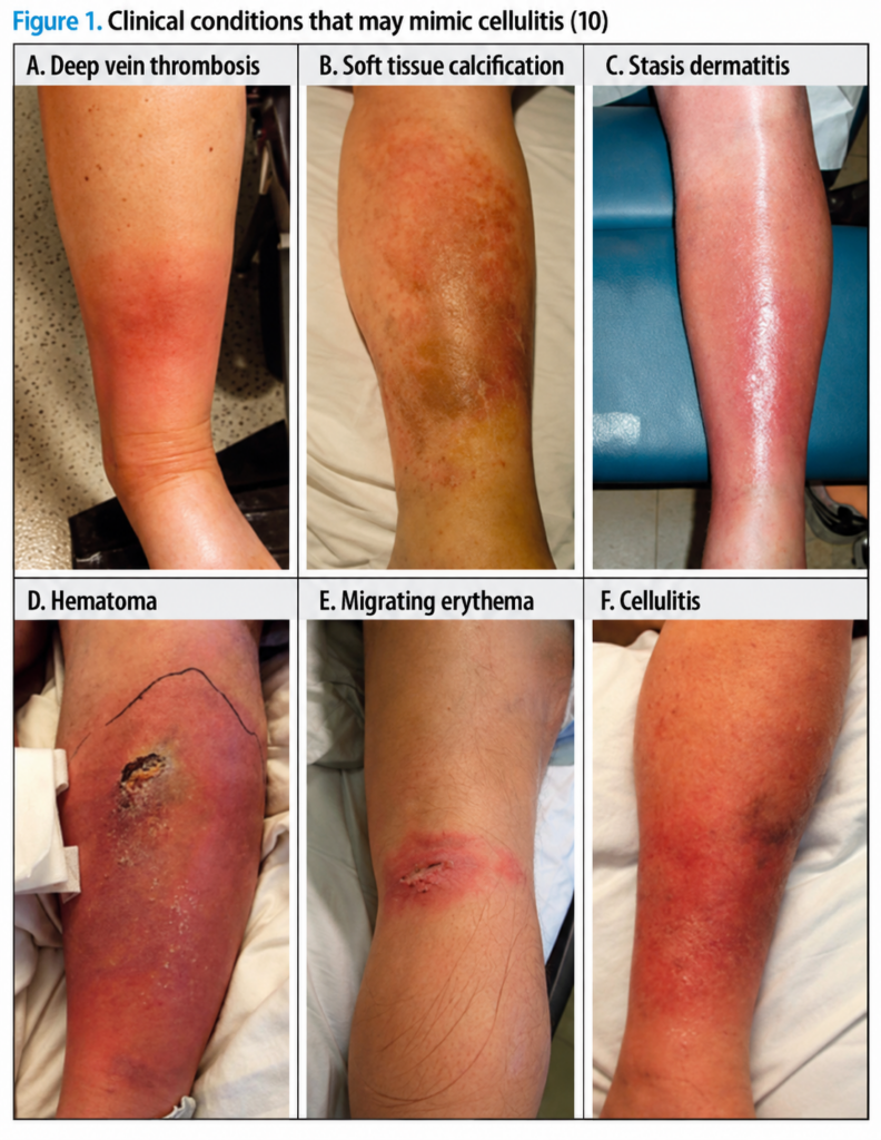
Pseudocellulitis may also present with fever, fatigue, or leukocytosis. It has been estimated that cellulitis is misdiagnosed in 33% of patients who are typically hospitalized because they fail to improve with standard outpatient treatment for cellulitis (20). The main conditions that should be differentiated from cellulitis include:
- Stasis dermatitis is the condition that most commonly resembles cellulitis (21). Stasis dermatitis is usually bilateral, whereas bilateral cellulitis is extremely rare in the setting of skin trauma (10). In rare cases, stasis dermatitis may be unilateral in patients who have sustained trauma to one leg or because of an anatomical variant (e.g., varicose veins).
- Hematoma is most commonly diagnosed in patients who have experienced trauma or are taking anticoagulants. The diagnosis of hematoma can be confirmed by ultrasonography.
- Gout may also present with fever and leukocytosis, while serum uric acid levels may not be elevated. Gout should be considered when erythema involves a joint. A positive response to nonsteroidal anti-inflammatory drugs and aspiration of synovial fluid may help differentiate gout from cellulitis (22, 23).
If there is no response to adequate treatment for cellulitis, or if the skin lesions are multiple, symmetrical, persistent, or slowly progressive, an alternative diagnosis should be considered (Table
Table 2. Differential Diagnosis of Cellulitis (22, 23)
| Category | Frequency | Conditions |
|---|---|---|
| Infectious | Common | Erythema migrans, Herpes simplex infection, Herpes zoster infection, abscess |
| Rare | Bacterial (erysipelas, necrotizing fasciitis), viral (parvovirus B19, CMV), fungal (Cryptococcus neoformans, Sporothrix schenckii, mucormycosis), mycobacterial infection, parasitic infections, osteomyelitis, septic arthritis | |
| Inflammatory | Common | Drug reaction, contact dermatitis, angioedema, Sweet syndrome, gout, acute bursitis, erythema nodosum |
| Rare | Pyoderma gangrenosum, sarcoidosis, eosinophilic cellulitis, relapsing polychondritis, polyarteritis nodosa, panniculitis | |
| Vascular | Common | Stasis dermatitis, lymphedema, deep vein thrombosis, superficial thrombophlebitis, hematoma |
| Rare | Erythromelalgia, soft-tissue calcification | |
| Neoplastic | Rare | Erysipeloid carcinoma, Paget disease, breast carcinoma, lymphoma, leukemia |
| Miscellaneous | Common | Insect bites, foreign-body reaction, subcutaneous injections |
| Rare | Compartment syndrome, pressure-induced blisters |
Treatment
The cellulitis treatment algorithm and antibiotic dose recommendations are based on the 2014 guidelines of the Infectious Diseases Society of America (17), the Johns Hopkins antibiotic guidelines, and the results of randomized controlled trials (24). The choice of antibacterial treatment for cellulitis depends on whether the infection is nonpurulent or purulent and on disease severity (mild, moderate, or severe). Therefore, antibiotic treatment recommendations for different forms of cellulitis are briefly reviewed below.
Typical Nonpurulent Cellulitis
Typical nonpurulent cellulitis without systemic signs of infection (mild cellulitis) should be treated with antibiotics active against streptococci (cephalexin, dicloxacillin, penicillin, amoxicillin/clavulanate); in patients allergic to penicillin-group antibiotics, clindamycin should be used (25).
In patients diagnosed with nonpurulent cellulitis who have at least one criterion of systemic inflammatory response syndrome (SIRS) (26), for example, temperature >38 °C or 90 beats/min, respiratory rate >20 breaths/min, leukocyte count >12,000/mm³ or
Purulent Cellulitis
In purulent cellulitis, the most likely causative organism is S. aureus (7). For purulent cellulitis without signs of systemic infection (mild cellulitis), when MRSA infection is not suspected, cephalexin or amoxicillin with clavulanic acid is prescribed, while clindamycin is used in patients allergic to penicillin.
If MRSA infection is suspected in mild purulent cellulitis, Biseptol or doxycycline should be prescribed. If both streptococcal and MRSA infection are suspected, cephalexin or penicillin should be added to Biseptol or doxycycline (24). For patients allergic to penicillin, clindamycin or linezolid is an alternative.
Patients diagnosed with purulent cellulitis who have one SIRS criterion (moderate cellulitis) may be treated in the same way as those with mild cellulitis. Patients with two or more SIRS criteria must be treated with intravenous antibiotics—oxacillin, nafcillin, or cefazolin if methicillin-sensitive S. aureus is suspected, or vancomycin, clindamycin, or linezolid if MRSA infection is suspected (29, 30).
In purulent cellulitis with SIRS criteria accompanied by hypotension, immunodeficiency, or rapid disease progression (severe cellulitis), empirical treatment with intravenous vancomycin, clindamycin, linezolid, daptomycin, or ceftaroline is recommended to provide antibacterial coverage against MRSA infection. The need for surgical treatment should also be considered because of a possible necrotizing process, and cultures of excised tissue should be obtained.
Empirical antibiotic use increases antimicrobial resistance among pathogens causing soft-tissue infections. The incidence of MRSA infection increased from 26.2% in 1998 to 47.4% in 2004. Empirical vancomycin use increased from 18% in 2000 to 69% in 2006. Low bactericidal activity and tissue penetration, as well as an insufficient drug dose or a prolonged course of treatment, have increased vancomycin resistance.
Because treatment failures with vancomycin have been observed and patients' body weight and body mass index are increasing, dosing of this drug should be based on the patient's weight (15–20 mg/kg intravenously every 8–12 hours) rather than on a standard dose of 1,000 mg every 12 hours. Vancomycin concentrations in the blood should be monitored because of the potential for nephrotoxicity (31, 32).
Once culture results are available, antibiotics should be changed to a narrower-spectrum agent. If there is no response to antibiotics within 24–48 hours, the diagnosis of cellulitis should be reassessed and the possibility of a resistant or atypical pathogen should be considered. In immunosuppressed patients, numerous microorganisms can cause cellulitis; therefore, in addition to bacteria, fungal, viral, and parasitic microorganisms should also be considered.
Duration of Treatment
The duration of treatment depends on the clinical response. Outpatient treatment lasts 5–10 days, while immunosuppressed patients are treated for 7–14 days (17). Pain, redness, swelling, and warmth at the affected site should be reassessed after 24–48 hours. If there is no improvement or the patient's condition deteriorates, antibacterial treatment should be changed or the possibility of a resistant microorganism or an alternative diagnosis should be considered (33).
Complications
Cellulitis requires treatment. There is a small but real risk of bacteremia, which occurs most commonly with streptococcal cellulitis. This may lead to complications such as endocarditis, glomerulonephritis, osteomyelitis, toxic shock, and elephantiasis. Cellulitis can damage the lymphatic vessels, and the resulting lymphedema acts as a predisposing factor for recurrent cellulitis. Cellulitis recurs in 22–49% of patients who have previously experienced one episode and usually occurs at the same site (6).
Prophylactic Treatment
The prophylactic use of antibiotics remains a matter of debate. It is thought that patients who experience cellulitis 3–4 times per year may be given prophylactic antibacterial treatment with oral penicillin 250 mg or 1 g twice daily, erythromycin 250 mg twice daily, clindamycin 150 mg once daily, or an intramuscular penicillin injection of 1.2 million IU for 4–52 weeks (34).
A double-blind, randomized controlled trial conducted in 2013 included 274 patients who had experienced two or more recurrent episodes of cellulitis. Some participants received penicillin 250 mg twice daily, while others received placebo. The study lasted 12 months. Antibacterial prophylaxis significantly reduced the risk of recurrent cellulitis of the legs, although the effect diminished after penicillin was discontinued. Several factors were found to potentially contribute to prophylaxis failure: body mass index ≥33 (p=0.01), three or more previous episodes of cellulitis (p<0.001), and existing edema (p=0.06) (34).
Summary
Cellulitis is a bacterial infection of the skin and subcutaneous tissues characterized by diffuse, poorly demarcated erythema, warmth at the affected site, swelling, and tenderness. Cellulitis is a common health problem worldwide. It is diagnosed on the basis of the patient's medical history and clinical presentation. Uncomplicated cellulitis is treated with antibiotics active against streptococci and methicillin-sensitive S. aureus. If treatment with first-line antibiotics is unsuccessful, resistant microorganisms, other diseases that may resemble cellulitis, or coexisting complicating conditions such as immunosuppression and chronic liver and kidney diseases should be considered.
Internistas (164)
References available from the editorial office.
