
Hyperprolactinemia: Diagnosis, Treatment and Monitoring
I
Introduction
Hyperprolactinemia is a condition in which the concentration of the hormone prolactin in the blood increases because of disturbances in its secretion or elimination. Prolactin is a polypeptide hormone produced by lactotroph cells and secreted by the anterior pituitary gland. Its secretion is not constant—higher concentrations are released during sleep, stress, pregnancy, or stimulation of the chest wall. Under physiological conditions, prolactin concentrations range from 212–424 mIU/L in men and from 212–530 mIU/L in women who are neither pregnant nor breastfeeding. Its half-life in the blood is 50 minutes (1).
The most important physiological function of prolactin is associated with lactation in women after pregnancy and childbirth. Prolactin promotes the development of milk-producing cells in the breast, activates milk production, and regulates its secretion. In addition, prolactin, also known as luteotropic hormone, suppresses the synthesis and secretion of gonadotropin-releasing hormones in the pituitary gland, leading to decreased synthesis of gonadotropins and sex hormones—estrogens in women and testosterone in men (1). In the absence of these hormones, women do not ovulate and experience menstrual cycle disturbances, while men develop impaired spermatogenesis, decreased libido, and reduced fertility. After childbirth, an increased prolactin concentration ensures successful lactation and protects women from another pregnancy. There is also evidence that prolactin influences the development of maternal behavior.
Prolactin secretion is regulated by various biologically active substances and neural stimulation (Figure 1):
- The constant concentration of prolactin in the blood is controlled by dopamine secreted in the pituitary gland, which regulates prolactin secretion through a positive feedback mechanism: as the prolactin concentration increases, dopamine secretion in the pituitary gland also increases. When dopamine reaches the pituitary gland, it binds to D2 receptors on lactotrophs and inhibits prolactin secretion. This maintains the physiological concentration of prolactin in the blood.
- The proliferation of prolactin-secreting cells in the pituitary gland is activated by estrogens, while secretion of the hormone itself is influenced by thyrotropin-releasing hormone (TRH) released by the pituitary gland. In clinical practice, its secretion is reflected by the TRH concentration, which should always be measured when hyperprolactinemia is diagnosed. An increased TRH concentration may be a cause of hyperprolactinemia.
When the need for prolactin increases, for example during breastfeeding, emotional and mechanical stimuli such as an infant sucking on the mother's breast effectively promote the secretion of this hormone, causing short-term activation of prolactin secretion lasting 30–45 minutes and ensuring lactation.
Figure 1. Regulation of prolactin metabolism
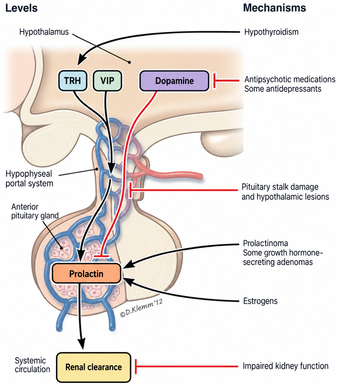
TRH – thyrotropin-releasing hormone; VIP – vasoactive intestinal peptides. Black arrows indicate activating factors, while red lines indicate inhibitory factors (2).
Hyperprolactinemia
Hyperprolactinemia is diagnosed three times more often in women than in men. This condition is most commonly diagnosed in patients aged 25–34 years, although in rare cases it may also develop in children and adolescents (3).
Because prolactin is produced by lactotroph cells of the pituitary gland, an increase in its concentration in the blood is usually caused by factors that lead to hyperplasia or hyperfunction of these cells, pituitary damage, or slowed hormone metabolism. These factors may be physiological, pathological, or pharmacological (Table 1).
Table 1. Causes of Hyperprolactinemia (4)
| Physiological Factors | Pathological Factors |
|---|---|
| Pregnancy, lactation | Systemic diseases |
| Chest wall stimulation | Chest wall pathology (injuries, surgeries, herpes zoster) |
| Sleep | Chronic kidney disease |
| Stress (physical and emotional) | Cirrhosis |
| Sexual intercourse | Epilepsy (with seizures) |
| Physical exercise | Polycystic ovary syndrome |
| Pharmacological Factors | Pituitary and hypothalamic lesions |
| Neuroleptics and antipsychotics | Tumors |
| Anesthetics | Injuries, surgeries |
| Antiepileptic drugs | Granulomas |
| Antidepressants | Infiltrations |
| Antihistamines | Hypersecretion of the pituitary |
| Antihypertensive drugs | Adenoma |
| Dopamine receptor blockers | Trauma, surgery |
| Dopamine synthesis inhibitors | Acromegaly |
| Opioid analgesics | Compressive tumors |
| Oral contraceptives (estrogens) | Lymphocytic hypophysitis |
Differential Diagnosis of the Main Causes of Hyperprolactinemia
When hyperprolactinemia is detected, the patient's subsequent treatment and care strategy depends on the underlying cause of the condition. To determine the origin of the increased prolactin concentration in the blood, it is important to:
- Collect a detailed patient history.
- Assess the possibility of pregnancy.
- Determine which medications the patient is taking and evaluate their influence on prolactin secretion.
- Pay attention to additional complaints, such as headaches or visual disturbances, which, together with a significantly elevated prolactin concentration of 10–20 times the normal level, may indicate a pituitary adenoma—prolactinoma (3).
- Evaluate concomitant diseases, paying particular attention to thyroid function. If hypothyroidism is detected, it should be treated with thyroxine. Kidney function should also be assessed, because in people with chronic kidney disease, the prolactin concentration may be elevated because of slower hormone elimination.
Hyperprolactinemia Caused by Pituitary Adenomas
Prolactin-secreting pituitary adenomas develop from lactotroph cells. These tumors account for up to 50% of all pituitary tumors and may be characterized by increased secretion not only of prolactin but also of other hormones, such as growth hormone, adrenocorticotropic hormone, and thyroid-stimulating hormone.
Depending on their size, prolactinomas are classified as microprolactinomas (5,300 mIU/L prolactin concentration), while macroadenomas are usually diagnosed in the presence of a significantly higher prolactin concentration (4). Microprolactinomas are more common in women than in men, with a ratio of 20:1, whereas the frequency of macroadenomas is the same in both groups, with a ratio of 1:1.
This distribution is explained by the fact that men have a longer asymptomatic period and delay seeking medical attention; therefore, by the time the diagnosis is established, the adenoma is more advanced. Most prolactinomas are benign, although in rare cases these tumors may metastasize.
Drug-Induced Hyperprolactinemia
A common cause of hyperprolactinemia is the use of medications, particularly neuroleptics and antipsychotics. Widely used antipsychotic drugs, including risperidone, phenothiazines, and haloperidol, are dopamine receptor antagonists. When these medications are used, the positive feedback control mechanism is suppressed, resulting in an increase in prolactin concentration in the blood (6). Unlike tumor-induced hyperprolactinemia, however, prolactin levels usually increase gradually and do not reach very high concentrations during treatment with these drugs (4).
The course of the disease in these patients is often asymptomatic, although specific clinical symptoms may occur. If hyperprolactinemia persists for a long period, bone density may decrease because of sex hormone deficiency. The diagnosis of drug-induced hyperprolactinemia can be confirmed by performing a drug withdrawal test. When the patient stops taking the suspected medication that may be causing the increase in prolactin, the prolactin concentration in the blood returns to normal within 3–5 days. If discontinuation of treatment is not possible, it is recommended to replace the medication with another drug that does not affect prolactin secretion. When changes to the patient's oral medication regimen are required, consultation with the treating psychiatrist is recommended (4).
Complaints and Symptoms
Women of reproductive age with elevated prolactin concentrations in the blood most commonly seek medical attention because of the classic triad of symptoms—amenorrhea or oligomenorrhea, infertility, and galactorrhea. Amenorrhea and infertility develop when elevated prolactin levels suppress gonadotropic hormone activity for a prolonged period. Galactorrhea is a direct manifestation of increased prolactin concentration because excess prolactin stimulates mammary gland activity.
Postmenopausal women typically have hypogonadism; therefore, hyperprolactinemia in this group does not usually present with the classic clinical symptoms. If hyperprolactinemia is caused by a pituitary macroadenoma, patients may seek medical attention only when symptoms caused by local tumor compression develop, such as headaches and visual disturbances. In some women, hyperprolactinemia may be detected incidentally.
Galactorrhea occurs less frequently in men because male breast tissue is less sensitive to prolactin than female breast tissue. Men most commonly present with fertility disorders, decreased libido, and oligospermia (1, 6).
Laboratory Diagnosis
Testing Prolactin Levels in the Blood
A single measurement of prolactin concentration in blood serum is considered sufficiently reliable and accurate for detecting hyperprolactinemia (4). If prolactin concentrations are found to be >424 mIU/L in men or >530 mIU/L in women in a sample taken at any time of day, hyperprolactinemia may be diagnosed.
If the result is close to the upper limit of the normal range or if the physician has other suspicions, the test should be repeated the following day, with several samples taken at intervals of 15–20 minutes (7, 8). It is essential that the patient prepares adequately for the test by controlling physiological factors: the patient should not have exercised, should be free from stress, should be well rested, and should not be taking medications that may affect prolactin secretion (4).
In patients with prolactinoma, a direct relationship between tumor size and serum prolactin concentration can often be identified. For example, patients with a prolactin concentration 10–20 times higher than normal are most commonly diagnosed with macroprolactinoma. Although this relationship is strong, some cases do not follow this pattern.
One of the most common reasons for such a discrepancy is the so-called hook effect, which occurs because of a laboratory testing artifact and results in the measurement of a low prolactin concentration despite an increased amount of prolactin in the blood. If a suspiciously low prolactin concentration is detected that does not correspond to the clinical presentation of the disease, it is recommended to repeat the test after diluting the blood serum at a ratio of 1:100 (4).
Testing for Macroprolactin Concentration in the Blood
In some patients, hyperprolactinemia is diagnosed incidentally in the absence of any clinical symptoms. In such cases, testing for macroprolactin is recommended. Although 85% of circulating prolactin in the blood is present in monomeric form, associated dimers—macroprolactin—are also present. The biological activity of this form of prolactin is low; therefore, patients with macroprolactinemia may have no complaints or symptoms (7, 8).
Radiological Diagnosis
When an elevated prolactin concentration is detected in the patient's blood serum, magnetic resonance imaging (MRI) of the head is recommended, except in cases where the patient is taking medications that can increase prolactin levels in the blood (4).
MRI may reveal functionally active adenomas in the pituitary gland whose increased secretion leads to hyperprolactinemia. It is important to assess the structure of the lesion. If the laboratory prolactin result is disproportionately low compared with the imaging findings, the patient may have a cystic pituitary adenoma in which most of the mass consists of functionally inactive tissue.
The size of the prolactinoma is also important when planning further treatment. In addition to prolactinomas, MRI may reveal other adjacent pathological lesions, such as cysts or tumors, which can increase hormone secretion through a mass or pressure effect on the pituitary gland.
Drug Treatment
Symptomatic hyperprolactinemia is currently treated with oral dopamine agonists—cabergoline, bromocriptine, quinagolide, and pergolide (9). Treatment begins with small doses that are gradually increased to the therapeutic dose.
When the treatment regimen is followed consistently, a rapid reduction in clinical symptoms may be observed. Visual symptoms regress within the first few days, while sexual function is restored within 2–3 weeks (10). If hyperprolactinemia is caused by an adenoma, a reduction in tumor size may be detected as early as 6 weeks after the start of treatment by repeating the MRI examination.
Cabergoline is currently the first-choice dopamine agonist because it is more effective and is associated with a lower risk of adverse effects than bromocriptine (4). This difference in effectiveness may be explained by the greater specificity of cabergoline for dopamine receptors on lactotroph cells. A lower likelihood of adverse effects may also improve adherence to the treatment regimen.
Adverse effects associated with dopamine agonists include nausea, postural hypotension, and impulsive behavior. Long-term use of high doses of cabergoline has been associated with fibrosis of the heart valves; therefore, periodic echocardiography is recommended (4).
Treatment and Long-Term Care for Drug-Induced Hyperprolactinemia
In patients with an asymptomatic course of the disease, treatment with dopamine agonists is not recommended (4). If the patient develops manifestations of prolonged hypogonadism, such as decreased bone density or other characteristic symptoms, replacement therapy with estrogen or testosterone is recommended.
Treatment of symptomatic hyperprolactinemia should begin with discontinuation of medications that affect prolactin concentration. If this is not possible, the medication should be replaced with another drug that has a similar therapeutic effect but does not cause hyperprolactinemia.
Specialists remain cautious about the use of dopamine agonists in such patients. Although these medications reduce prolactin levels in 75% of patients, clinical studies have shown that their use may exacerbate psychotic states (11).
Treatment and Long-Term Care for Adenoma (Prolactinoma)-Induced Hyperprolactinemia
Of all pituitary tumors, prolactinomas respond well to pharmacological treatment. Dopamine agonists normalize laboratory parameters, reduce tumor mass, and eliminate clinical symptoms.
- In patients with microprolactinoma who have no clinical symptoms of hyperprolactinemia and are not planning to have children, treatment with dopamine agonists is not recommended. These patients should be monitored, prolactin concentrations in the blood should be measured periodically, and MRI should be repeated if prolactin concentrations increase significantly or clinical symptoms develop (4).
- Dopamine agonists are recommended for symptomatic adenomas of any size. According to various clinical studies, these drugs reduce adenoma size, eliminate pre-existing visual disturbances and galactorrhea, restore fertility, and normalize prolactin concentrations in blood serum.
- Patients receiving pharmacological treatment require periodic monitoring. The course of clinical symptoms should be assessed and prolactin concentrations in blood serum should be measured, beginning 1 month after the start of treatment and continuing until prolactin concentrations normalize. After 1 year, MRI should be repeated. It may be performed after 3 months if macroprolactinoma has been diagnosed, if prolactin concentrations continue to increase despite treatment, or if new clinical symptoms develop.
- If a patient has been treated with dopamine agonists for at least 2 years, has a normal prolactin concentration in the blood, and has no signs of adenoma on MRI, discontinuation of treatment may be considered. Active monitoring should then be continued. Prolactin should be measured every 3 months during the first year and annually thereafter. If prolactin concentrations increase, MRI should be repeated. Studies have shown that the risk of prolactinoma recurrence is highest during the first year after discontinuation of treatment and is directly related to the previous size of the adenoma (4, 12).
Treatment of Drug-Resistant Prolactinomas and Long-Term Care
Most patients treated with standard doses of dopamine agonists experience a positive effect: prolactin concentrations in the blood normalize and the adenoma decreases in size. However, in some cases the response to treatment is insufficient—prolactin concentrations remain elevated, the adenoma decreases by less than 50%, or fertility disorders persist (14). These patients are diagnosed with resistance to dopamine agonists (13).
In some cases, the response to treatment may be contradictory: the adenoma decreases in size by more than 50%, but prolactin levels remain elevated, or vice versa. Resistance is more common in macroprolactinomas, occurring in 18% of cases, than in microprolactinomas, where it occurs in 10% of cases (4). This phenomenon has been observed more frequently in men than in women.
It is important not to confuse drug resistance with drug intolerance. Drug intolerance occurs when the adverse effects of medication are more severe and more numerous than the therapeutic benefits. In patients who are resistant to medication, the dose of dopamine agonists should be increased gradually according to changes in blood prolactin concentrations, up to the maximum tolerated therapeutic dose.
Transsphenoidal pituitary surgery may be recommended for patients with prolactinoma who do not tolerate medical treatment or in whom treatment remains ineffective even when high doses are prescribed. After surgery, the probability of prolactinoma recurrence ranges from 7% to 50% (15).
Summary
Hyperprolactinemia is a hormonal disorder that causes specific symptoms and may be encountered in the practice of family physicians, obstetrician-gynecologists, endocrinologists, and other specialists. Simple laboratory diagnosis of this syndrome and accessible treatment methods make it possible to correct the condition and maintain a stable patient status relatively easily.
When treatment recommendations are followed, the prognosis is generally good in many cases, and the disease does not have a negative impact on the patient's health, ability to work, or physical activity.
Publication "Internistas" No. 2, 2018
References: 1. Kasper D, et al. Harrison‘s Principles of Internal Medicine, 19th Ed. New York: McGraw Hill; 2015. 2. Huang W, Molitch ME. Evaluation and management of Galactorrhea. Am Fam Physician, 2012 Jun 1;85(11):1073-1080. 3. Kars M, et al. 2009 Estimated age- and sex-specific incidence and prevalence of dopamine agonist-treated hyperprolactinemia. J Clin Endocrinol Metab 94:2729–2734. 4. Melmed S, et al. Diagnosis and Treatment of Hyperprolactinemia: An Endocrine Society Clinical Practise Guideline, 2011. The Journal of Clinical Endocrinology & Metabolism, 96;2: 273–288. 5. Klibanski A. 2010 Clinical practice. Prolactinomas. N Engl J Med 362:1219-1226. 6. Melmed S, Kleinberg D 2008 Anterior pituitary. In: Kronenberg HM, Melmed S, Polonsky KS, Larsen PR eds. Williams textbook of endocrinology. 11th ed. Philadelphia: Saunders Elsevier. 7. Casanueva FF, et al. 2006 Guidelines of the Pituitary Society for the diagnosis and management of prolactinomas. Clin Endocrinol (Oxf) 65:265–273. 8. Mancini T, Casanueva FF, Giustina A. 2008 Hyperprolactinemia and prolactinomas. Endocrinol Metab Clin North Am 37:67–99. 9. Kearns AE, et al. 2000 Risperidone-associated hyperprolactinemia. Endocr Pract 6:425–429 10. Pollock A, McLaren EH 1998 Serum prolactin concentration in patients taking neuroleptic drugs. Clin Endocrinol (Oxf) 49:513–516. 11. Cavallaro R, et al. 2004 Cabergoline treatment of risperidone-induced hyperprolactinemia: a pilot study. J Clin Psychiatry 65:187–190. 12. Dekkers OM, et al. 2010 Recurrence of hyperprolactinemia after withdrawal of dopamine agonists: systematic review and meta-analysis. J Clin Endocrinol Metab 95:43–51. 13. Molitch ME 2003 Dopamine resistance of prolactinomas. Pituitary 6:19–27. 14. Gillam MP, et al. 2006 Advances in the treatment of prolactinomas. Endocr Rev 27:485–534. 15. Losa M, et al. 2002 Surgical treatment of prolactin-secreting pituitary adenomas: early results and long-term outcome. J Clin Endocrinol Metab 87:3180–3186.